
Essential Knowledge About Pain
“My scan doesn’t dictate my outcome”
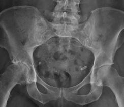
X-RAY
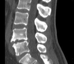
CT Scan
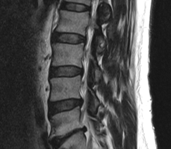
MRI
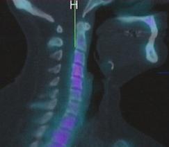
Bone Scan
Important Fact:
Only 10% of musculoskeletal injuries have findings on scans that are relevant. That means 90% do not, despite common held beliefs or sensations that “something must be ‘wrong’ inside my body that a scan can identify”.
Skilled clinical examination is required to match scan results with the clinical presentation to identify the 10% of people where scans do explain the symptoms.
A lot of time can be wasted searching for a specific cause of pain on scans, when one often does not exist. This does not mean that you don’t have pain, it just means that the scans are clear or findings are not related to the pain you are experiencing. This is the case 90% of the time.
WHEN DO I NEED A SCAN?
There are specific clinical indications for when scans are needed, and for what types of scans should be performed. Your health care practitioners will know the rules for when scans are necessary.
Important Fact:
Findings on scans are extremely common in individuals without pain, even in young people.
Ultrasound

EMG/Nerve Conduction

Blood Tests
Important Fact:
There is no correlation of scan findings with pain, disability, or ability to work.
Important Fact:
The way scans are reported and understood can have a significant negative effect on people. Words and phrases like ‘wear and tear’, ‘degeneration’, ‘narrowing’, ‘trapped’, ‘bulging’ and ‘protrusion’ in scan reports can be interpreted in a negative way by patients. When this occurs it has been associated with unhelpful beliefs and worse outcomes. Understanding that many of these changes occur in pain free individuals and/or are not related to your symptoms can be a helpful message. Your health care practitioner will be able to interpret the meaning of scans based on your clinical findings.
Intersting Fact:
One study followed over 600 people who had early MRI’s in their injury, versus 2400 who did not. Those who had early scans did worse than those who did not. Even those where guidelines suggested they should have scans did worse than those with the same problem but didn’t end up getting the scans. These results highlight the negative effect that scans can have on those with injuries. Also scans can result in unhelpful treatment practices.
Intersting Fact:
Scans results often don’t change after pain subsides and normal function is regained. A good example of this is MRI scan on athletes who have suffered a hamstring injury, gone through rehabilitation and successfully returned to sport. 90% still have changes on MRI despite returning to sport.
Summary: Scans don’t dictate outcome
The majority of the time, scans do not explain pain and disability. Incidental findings are extremely common. Poorly reported and understood findings can have a negative effect on people. Your scans do not dictate what you can achieve. Your health care practitioners can help you determine the relevance and meaning of scans.
Evidence Informed Information Compiled By Dr Darren Beales, PhD and Dr Tim Mitchell, PhD